Chiari Malformation
Chiari Malformation Treatment in Newport Beach
Chiari Malformation is an uncommon disorder that occurs when the skull is atypically small in size or deformed causing the brain tissue to move into the spinal canal. Chiari malformation type I occurs as the brain and skull are still growing, thus detection may not happen until late adolescent to early adulthood. Chiari malformation type II is a congenital disorder and the most common type of the two.
Chiari malformation interrupts the flow of cerebrospinal fluid that protects your brain and spinal cord. It can also cause an interference of signal transmission.
[fusion_builder_container hundred_percent=”yes” overflow=”visible”][fusion_builder_row][fusion_builder_column type=”1_1″ background_position=”left top” background_color=”” border_size=”” border_color=”” border_style=”solid” spacing=”yes” background_image=”” background_repeat=”no-repeat” padding=”” margin_top=”0px” margin_bottom=”0px” class=”” id=”” animation_type=”” animation_speed=”0.3″ animation_direction=”left” hide_on_mobile=”no” center_content=”no” min_height=”none”]
Chiari malformation type I occurs when the part of the skull housing the cerebellum is mishapen or too small leading to the tissue in the lower part of the cerebellum to move in to the spinal cord.
Chiari malformation type II is typically linked with myelomeningocele, a form of spina bifida, in which the backbone and the spinal canal haven’t closed properly before birth.
Most sufferers of chiari malformation have vague or no symptoms altogether. Commonly chiari malformation is detected only when tests are performed for other disorders. However, depending on the type and severity, chiari malformation can a variety of problems.
Chiari Malformation Type I
People with Chiari malformation type I can show the following symptoms:
- Numbness
- Problems with balance and coordination
- Muscle weakness
- Vision problems
- Headaches
- Dizziness
Chiari Malformation Type II
With chiari malformation type II, more tissue extends into the spinal canal and the symptoms can include those related to myelomeningocele.
- Changes in breathing pattern
- Severe headache
- Quick downward eye movements
- Weakness in arms
- Swallowing problems, such as gagging
- Numbness
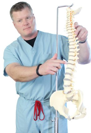
To diagnose chiari malformation, your Newport Beach orthopaedic surgeon, Dr. Todd Peters will perform a complete and thorough physical examination as well as a medical history check. He may also request X-Rays, MRIs, and CT scans to better assess your case.
X-Rays – A type of quick imaging process using electromagnectic waves to better show Dr. Peters bones of the body to pinpoint possible sources of pain.
Magnetic Resonance Imaging (MRI) – This type of imaging uses radiology to show Dr. Peters the soft tissues of the body like muscles, disks, nerves, and the spinal cord.
Computerized tomography (CT) – CT scans are X-ray scans taken from various angles to create a cross-section of bones and soft tissues of the body.
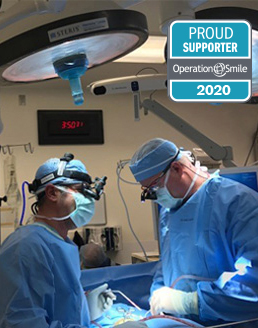
If sever enough, Dr. Peters may also recommend surgery to alleviate the pressure and regulate the progression of chiari malformation.
Dr. Todd Peters specializes in minimally invasive orthopaedic surgery to treat chiari malformation. For appointments, please call (949) 383-4182 or Contact Us.[/fusion_builder_column][/fusion_builder_row][/fusion_builder_container]